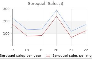
"Order 100 mg seroquel, medications zetia". V. Jarock, M.B. B.CH. B.A.O., M.B.B.Ch., Ph.D. Deputy Director, University of Cincinnati College of Medicine
In these midline regions treatment juvenile arthritis cheap 100 mg seroquel with visa, ruling out the presence of histone h3 K27M mutation is essential medicine 219 order seroquel 200 mg online. A classic radiologic (and macroscopic) presentation is the "butterfly" sample as a end result of symptoms 4 weeks pregnant generic seroquel 200 mg spread of the tumor across the corpus callosum to the contralateral hemisphere symptoms 5th disease cheap seroquel 100 mg overnight delivery. In the tissue surrounding the tumor, there may be considerable vasogenic edema manifested on imaging studies as hyperintensity on a T2-weighted MrI scan. A "variegated" appearance with strong gray-pink tissue on the periphery and yellow zones of central necrosis is typical (fig. As such sufferers could present with a number of foci seemingly distant from main tumor mass, creating the impression of a multifocal lesion on neuroimaging (see discussion 26 � of gliomatosis cerebri additional within the chapter). While true multifocal gliomas could occur, their exact frequency appears to be a lot decrease than their previously estimated range (2. Multifocal tumors would by definition be polyclonal and confirmed so solely by molecular evaluation. In some cases, the tumor cells might have considerable nuclear and cytoplasmic pleomorphism with multinucleated large cells (fig. Microscopic options: (B) mobile anaplasia, mitoses (h&E); (C) necrosis with pseudopalisading (h&E); (D) microvascular proliferation with glomeruloid structures (h&E). Microvascular proliferation is incessantly related to thrombosis of the affected vessels. Glioblastoma is among the most wellcharacterized cancers at the molecular level and was one of the first cancers to be studied utilizing large-scale integrative genomic approaches. The h3 K27M mutation is mostly present in midline tumors of the pons and thalamus, and the h3 G34r/V mutation is typically current in tumors arising in the cerebral hemispheres. Tumors which have h3 mutations and are in the midline structures (thalamus, brainstem, spine) are referred to in the WhO 2016 classification as a separate class, designated diffuse midline gliomas, h3 K27M mutant. Affected sufferers typically have a poor prognosis, although some stories have advised a somewhat higher clinical consequence (>10% 5-year survival), which if true could probably end result from higher surgical access to a extra circumscribed tumor mass. The tumor is often situated in the cerebral hemispheres, most frequently involving the temporal lobes. The sarcomatous regions normally include malignant spindle cells organized in a fascicular, herringbone, or typically a "storiform" pattern and should not often present different forms of mesenchymal differentiation, including cartilage, bone, skeletal, and smooth muscle. There appears to be a monoclonal origin of each the glial and mesenchymal components of the tumor, with the mesenchymal component most likely representing a complicated type of glial-mesenchymal transition analogous to epithelial-mesenchymal transition in carcinomas. The designation oligodendroglioma applies to a diffusely infiltrating glioma composed of tumor cells morphologically resembling mature oligodendrocytes. The tumor accounts for roughly 5% of all intracranial gliomas and happens mostly in adults, with a peak incidence between the ages of 30 and 60. The tumor must be distinguished from a separate category of diffuse leptomeningeal glioneuronal tumors which will additionally demonstrate "oligodendroglioma" morphologic elements together with the 1p/19q co-deletion. Because of the slowgrowing nature of the tumor, patients might have a long history of neurological symptoms, most frequently a seizure dysfunction. Imaging research usually show a well-demarcated mass, usually with calcification and occasionally intratumoral hemorrhage; peritumoral edema and contrast enhancement are unusual. The macroscopic look of oligodendroglioma is characteristically that of a variably wellcircumscribed, grayish pink neoplasm together with areas of mucoid change, which may lead to a gelatinous consistency as properly as zones of cystic degeneration, focal hemorrhage, and calcification. In routine paraffinembedded sections stained with h&E, the tumor cells are closely packed and appear swollen, consisting of a small, spherical nucleus (usually barely bigger than a traditional oligodendrocyte) surrounded by a transparent halo (fig. The tumor stroma also characteristically contains a network of thin-walled branching capillaries, typically described as a "chicken-wire" or "wishbone" vascular branching sample (fig. Anaplastic oligodendroglioma: (D) microvascular proliferation (h&E); (E) nuclear atypia, minigemistocytes (h&E). In some cases, the tumor could comprise nodules of increased cellularity; careful attention to these areas may reveal other anaplastic features inside them. Small calcifications (calcospherites) are a attribute histologic feature (fig. Another attribute and diagnostically useful characteristic is the presence of perineuronal, perivascular, or subpial tumor aggregates when the tumor infiltrates cortex. They are thought to be both a transitional kind between oligodendrocytes and astrocytes or a phenotypic recapitulation of the premyelination stage of regular immature oligodendrocytes or their progenitors. Concurrent whole-arm co-deletion of 1p and 19q, ensuing from a translocation involving these chromosomes, is integral to the analysis of oligodendroglioma. Some patients, however, develop multiple postoperative recurrences with eventual progression to frank anaplasia. The macroscopic appearance is just like oligodendroglioma besides that areas of necrosis may be present. The pilocytic astrocytoma (WhO grade I) tumor in its basic kind is properly circumscribed, sluggish growing, and infrequently cystic and predominantly happens in children and young adults (fig. The most common sites of incidence are the cerebellum and cerebral midline buildings, corresponding to hypothalamus/third-ventricular area, optic nerves, or brainstem, however the tumor can also originate in the cerebral hemispheres and spinal wire however usually with much less basic histological features than seen in the cerebellum. Imaging studies show a circumscribed, cystic, or (less often) strong mass with distinction enhancement in most all circumstances. In the cystic examples, distinction enhancement could also be localized to a localized mass of tumor throughout the cyst wall ("mural nodule"). The microscopic options of pilocytic astrocytoma are highly distinctive in their traditional form inside the posterior fossa tumors. The proportion of these two attribute patterns may range within a given tumor, and a few instances (especially when only a small biopsy amount is out there for study) might present a predominance of one pattern. In pilocytic astrocytoma, microscopic examination may reveal options that might be mistaken as proof of anaplasia; these embrace frequent definitive microvascular proliferation (including glomeruloid vessels), nuclear atypia, extension into the subarachnoid house, and occasionally bland (infarct-like) areas of necrosis. In most patients with pilocytic astrocytoma, the scientific course is indolent; the treatment of selection is surgical excision, with glorious long-term survival. Malignant transformation of a pilocytic astrocytoma could be very rare, and most case stories have occurred in patients following radiotherapy. Such alterations can be identified in more than 90% of cerebellar pilocytic astrocytomas but is discovered to a lesser diploma in gliomas with piloid features in the supratentorial areas (~50%). Notably, pilocytic astrocytoma inside the optic nerve is the brain tumor mostly present in patients with in neurofibromatosis type 1, a dysfunction that happens in roughly 1 in 3000 individuals. Pilomyxoid astrocytoma is a variant of pilocytic astrocytoma that most usually localizes to the hypothalamic region as a strong, well-circumscribed, contrast-enhancing mass. Almost all of those tumors are situated above the tentorium and with a preponderance originating within the temporal lobes. These low-grade features stand in marked contrast to the alarming cytologic pleomorphism. Some examples also categorical Chapter 2 Tumors of the Central Nervous System � 33 "neuronal" markers, together with synaptophysin and neurofilament protein. Other attribute however not invariable histologic options include the presence of a conspicuous reticulin network (possibly reflecting a putative origin from subpial astrocytes), lymphocytic infiltrates, and eosinophilic granular bodies similar to those found in ganglion cell tumors. This tumor is characteristically associated with tuberous sclerosis syndrome, occurring in as much as 20% of sufferers with this genetic dysfunction. It stays unresolved whether the tumor can occur in sufferers with out tuberous sclerosis. Most circumstances come up through the first 20 years of life, and sufferers present with worsening of a seizure disorder or signs of increased intracranial stress. The tumor consists of relatively large cells resembling gemistocytic astrocytes but typically having "ganglioid" nuclei with outstanding nucleoli (fig. Immunohistochemically, the tumor cells could express both or each glial and neuronalassociated antigens, which can replicate their putative origin from dysplastic bipotential cells in the subependymal region. Although the tumor impacts individuals in all age teams, it happens extra regularly in childhood and adolescence. Any degree of the ventricular system could be the primary web site of origin; nonetheless, the most typical website is the posterior fossa across the fourth ventricle (approximately 60%). In the spinal twine, ependymomas are the most typical neuroepithelial tumor, accounting for about 60% of spinal gliomas. Imaging research show a comparatively well-circumscribed mass with various degrees of contrast enhancement; tumor infiltration and edema are relatively rare. Macroscopically, the tumor is usually a gray-red, lobulated, and often well-demarcated mass near a ventricular cavity (fig. Some infratentorial tumors might lengthen into the cerebellopontine angle or inside the cisterna magna along the medulla. Chapter 2 Tumors of the Central Nervous System � 35 the looks of an ependymoma in the spinal wire is that of a well-circumscribed intramedullary mass displacing normal structures.

Clinicians should be familiar with these imaging findings to keep away from diagnosing this mass as a more ominous tumor similar to a liposarcoma treatment canker sore 200 mg seroquel discount with amex. A symptoms 5 months pregnant cheap 200 mg seroquel with amex, Coronal T1-weighted picture reveals a fusiform mass with signal intensity isointense to skeletal muscle (arrows) treatment for plantar fasciitis seroquel 100 mg buy overnight delivery. B symptoms bipolar disorder 50 mg seroquel discount overnight delivery, Coronal T2-weighted picture exhibits heterogeneously hyperintense sign throughout the mass with an elongated tail distally, suggestive of neurogenic etiology. C, Postcontrast T1-weighted fat-saturated picture shows thick nodular enhancement of periphery with lack of central enhancement, suggestive of necrosis. Coronal T1-weighted image (A) reveals giant delicate tissue mass centered round distal two-thirds of first metacarpal with isointense signal to adjoining muscle. T2-weighted coronal (B) and axial (C) pictures present heterogeneously elevated T2 sign and destruction of first metacarpal and phalanges. A, Axial T1-weighted picture demonstrates massive mass in posteromedial thigh abutting posterior cortex of distal femur. Axial T1-weighted (A) and T2-weighted (B) photographs present a mass to inferior border of the scapula with sign intensity much like muscle. C, T1-weighted fat-saturated postcontrast picture exhibits mild enhancement of this mass. A, Axial T1-weighted picture reveals massive masslike collection in medial thigh with areas of hyperintense sign, suggestive of subacute blood merchandise (arrows). B, T2-weighted axial image exhibits hypointense rim that can additionally be suggestive of blood products. C, Postcontrast T1-weighted fat-saturated picture reveals minimal peripheral enhancement, typical of a hematoma. Sagittal (A) and axial (B) T2-weighted pictures show intraarticular synovial-based mass with predominantly hypointense signal intensity (arrows). Sagittal (A) and axial (B) T2-weighted images show synovial our bodies especially around posterior aspect of joint (arrows). Hyperintense T2 sign within synovial bodies is typical of noncalcified synovial chondromatosis. Coronal T1-weighted (A) and T2-weighted (B) images show a group associated with tibiofibular joint extending into adjoining delicate tissues (arrows). C, Postcontrast T1-weighted fat-saturated coronal picture exhibits minimal peripheral enhancement of this assortment. C, Postcontrast T1-weighted fat-saturated image exhibits minimal peripheral enhancement of the mass, typical of a ganglion or synovial cyst (arrows). Coronal (A), sagittal (B), and axial (C) T2-weighted photographs of left shoulder show intramuscular fluid collection involving supraspinatus tendon (arrows). On A, narrow neck of fluid extends into tendon floor (arrow), according to partial tear. Sagittal (A) and coronal (B) proton-density-weighted photographs of left knee show focal fluid accumulation in semimembranosus�medial head gastrocnemius bursa according to a popliteal cyst (arrows). C, Axial T2-weighted picture exhibits semimembranosus tendon and medial head gastrocnemius tendon anterior to popliteal cyst (arrows). Mild peripheral enhancement is famous within the postcontrast T1-weighted fat suppressed images (C). A, T2-weighted sagittal picture of right arm reveals large intramuscular collection with surrounding reactive edema. B, T1-weighted fat-saturated postcontrast picture exhibits nodular enhancement in periphery of the collection (arrows), typical of an abscess. Initial coronal T1-weighted (A) and T2-weighted (B) pictures by way of proper shoulder show complex-appearing soft tissue mass with elevated T2 sign intensity and surrounding muscle edema (arrows). Axial T2-weighted image (A) and axial T1-weighted fat-saturated postcontrast image (B) of the pelvis present giant collections round hip joints bilaterally with peripheral enhancement and diffuse, surrounding delicate tissue edema, suggestive of a nonossified matrix. T1-weighted sagittal (A) and axial (B) photographs of proper ankle show a flexor digitorum accessorius muscle in region of tarsal tunnel (arrows). C, Proton-density-weighted axial image exhibits anterior displacement of neurovascular bundle on account of mass effect of this accessory muscle. Coronal T1-weighted (A) and sagittal T2-weighted (B) images present hypointense mass overlying the greater trochanter (arrows). C, Axial postcontrast T1-weighted fat-saturated image reveals peripheral enhancement of this mass (arrows). Axial T1-weighted (A) and T2-weighted (C) images present small mass with poorly defined margins on ulnar aspect of distal forearm abutting the fascia (arrows). B, Postcontrast T1-weighted fat-saturated picture reveals mild to average enhancement (arrows). Axial T2-weighted photographs with out (A) and with (B) fat suppression show a fatty mass associated with the ulnar nerve (arrows). A, T1-weighted sagittal image of the left leg shows a pretibial mass (arrows) with hypointense signal and poorly defined margins. There is related pores and skin thickening and edema, typical of huge localized lymphedema. Classification and management of the varied superficial vascular anomalies: hemangiomas and vascular malformations. Current ideas in the classification, diagnosis and remedy of vascular anomalies. Granular cell tumor of the extremity: magnetic resonance imaging traits with pathologic correlation. Variability in the presentation of synovial sarcoma in youngsters: a plea for higher consciousness. Epithelioid sarcoma: the clinicopathological complexities of this rare gentle tissue sarcoma. Pigmented villonodular synovitis of synovial joints: scientific, pathologic, and radiologic features. Pigmented villonodular synovitis and big cell tumors of the tendon sheath: radiologic and pathologic options. Lipofibromatous hamartoma of the upper extremity: a evaluate of the radiologic findings for 15 patients. Massive localized lymphedema: a clinicopathologic research of 46 sufferers with an enrichment for multiplicity. The types and patterns of mutation are highly variable and heterogeneous amongst, and generally inside, different tumor entities, starting from ploidy shifts to single-base substitutions and epigenetic alterations. The latter forms of mutation, showing a powerful association with a specific tumor phenotype, can be exploited for scientific diagnostic purposes. Indeed, genetic analyses are now routinely used as an adjunct to traditional morphologic and immunohistochemical investigations within the diagnosis of many neoplasms, including soft tissue tumors. It is also well acknowledged that certain forms of mutation have a powerful influence on the aggressiveness of the tumor cells, and that the mutation standing thus should be assessed to select the right sort and degree of therapy. Furthermore, numerous novel treatment methods have just lately been developed, usually with a dramatic enchancment of affected person consequence, based on the identification of specific mutations in tumor cells, similar to kinase inhibitors for chronic myeloid leukemia, malignant melanoma, or gastrointestinal stromal tumor. Similarly, genetic analyses have demonstrated that the clinical and biologic variation amongst delicate tissue tumors is mirrored in their genotypes, and it was just lately shown that the addition of genetic info considerably improves the diagnostic precision. Thus the use of supportive genetic information varies considerably amongst sarcoma centers, relying on native traditions, technical and economic situations, and ability of the pathologists. The cursory knowledge of only some gentle tissue tumors however, many genetic options have been shown to be strongly related to morphologic options, and a rapidly growing subset of mutations promise to shed light on affected person end result. This article discusses major molecular pathogenetic options of soppy tissue tumors, specializing in those that are clinically relevant, both as diagnostic markers or for treatment stratification. In addition, the expression or operate of proteins may be affected by roughly secure epigenetic adjustments, similar to histone methylation, in addition to by posttranslational modifications, similar to glycosylation of proteins. Consequently, mutations that are associated with neoplastic transformation range from single nucleotide variants to complex genomic modifications affecting entire chromosomes, augmented by epigenetic changes. Several such mutations are known to be associated with an increased risk of creating gentle tissue tumors (Table four. Often the phenotypic consequences of these mutations are quite extensive, leading to a recognizable collection of phenotypic effects-a syndrome-that might embody malformations and intellectual impairment, as properly as an increased threat for varied neoplasms. Although inherited most cancers predisposition is often caused by small genetic variants, constitutional chromosomal rearrangements may also occasionally lead to an elevated threat for gentle tissue tumors. However, as a outcome of most predisposing syndromes are exceedingly uncommon, and gentle tissue tumors could appear anyplace in the body, no clinical consensus has been reached regarding if and the way the patients and their family members must be followed with regard to sarcomas. Therefore an prolonged investigation of the family, beginning with the parents, is warranted to identify siblings and different family members who may be carriers of the identical mutation and thus ought to be monitored for early cancer detection.
The cells of myxoid leiomyoma are larger and have more plentiful eosinophilic cytoplasm than those of aggressive angiomyxoma administering medications 7th edition ebook 100 mg seroquel purchase with mastercard, and have a tendency to be arranged in small packets or free fascicles medicine numbers 100 mg seroquel purchase otc. Careful inspection medicine 5852 200 mg seroquel generic visa, nonetheless symptoms 5 weeks pregnant buy discount seroquel 300 mg on-line, will invariably reveal extra typical zones of fibromatosis, with lengthy, sweeping fascicles of uniform myofibroblastic cells arrayed about a thin-walled, dilated vasculature, often displaying perivascular edema. Myxofibrosarcoma shows a a lot greater diploma of cytologic atypia than deep angiomyxoma and a different vascular pattern, with thick-walled arborizing vessels from which the neoplastic cells appear to emanate. Fibroepithelial stromal polyps of the vulvovaginal area have a wide spectrum of morphologic appearances and thus enter into the differential diagnosis of the varied genital stromal tumors. Fibroepithelial stromal polyps usually come up in younger to middle-aged girls, usually within the vagina. Histologically, this lesion exhibits a distinctly polypoid development sample and lacks a grenz zone with the overlying mucosa or skin. The absence of this grenz zone is in distinction to true genital stromal tumors and may be a valuable clue to the correct prognosis. Fibroepithelial stromal polyps show vital variability from case to case, with some being hypocellular and myxoid and others showing marked hypercellularity with a pseudosarcomatous look. There is great immunophenotypic overlap with the varied genital stromal tumors as a end result of the stromal cells of fibroepithelial polyps may categorical desmin, actin, estrogen, and progesterone receptors. The clinical appearance and site of the lesions usually counsel the right analysis. The round ligament, when removed incidentally during repair of an inguinal hernia, can also be misinterpreted as a leiomyoma. B, Microscopic part exhibits well-differentiated clean fibers oriented perpendicular to the skin floor. Smooth muscle tumours of the external genitalia: clinicopathological evaluation of a collection. Fumarate hydratase mutations and predisposition to cutaneous leiomyomas, uterine leiomyomas and renal most cancers. Clinical features of a number of cutaneous and uterine leiomyomatosis: an underdiagnosed tumor syndrome. The problem in predicting habits of smooth-muscle tumors in deep gentle tissue. An analysis of highly differentiated smooth muscle tumors of deep soft tissue supporting two distinct subtypes. Retroperitoneal leiomyomas: a clinicopathologic and immunohistochemical examine of fifty six cases with a comparability to retroperitoneal leiomyosarcomas. Studies of cell floor and connections in normal and achalasia esophageal easy muscle. The ultrastructure of smooth muscle tumors with a consideration of the attainable relationship of glomangiomas, hemangiopericytomas, and cardiac myxomas. A monoclonal antibody towards alpha-smooth muscle actin: a brand new probe for clean muscle differentiation. Inguinal easy muscle tumors in girls: a dichotomous group consisting of m�llerian-type leiomyomas and soft tissue leiomyosarcomas-an analysis of fifty five instances. Multiple peritoneal leiomyomas related to a granulosa-cell tumor of the ovary. Peritoneal leiomyomatosis (leiomyomatosis peritonealis disseminata): a clinicopathologic study of 20 cases with ultrastructural observations. Histogenesis of leiomyomatosis peritonealis disseminata (disseminated fibrosing deciduosis). Ultrastructure of myofibroblasts and decidualized cells in leiomyomatosis peritonealis disseminata. Benign glandular inclusion of the peritoneum related to leiomyomatosis peritonealis disseminata. Sex cord�like sample leiomyomatosis peritonealis disseminata: a hitherto undescribed function. Disseminated peritoneal leiomyomatosis: clonality evaluation by X chromosome inactivation and cytogenetics of a clinically benign easy muscle proliferation. Adjuvant surgical and hormonal treatment of leiomyomatosis peritonealis disseminata: a case report. Computed tomography of leiomyomatosis peritonealis disseminata with malignant transformation. Angiomyofibroblastoma of the vulva: a benign neoplasm distinct from aggressive angiomyxoma. Aggressive angiomyxoma: reappraisal of its relationship to angiomyofibroblastoma in a series of 16 cases. Angiomyofibroblastomalike tumor of the male genital tract: analysis of eleven instances with comparability to feminine angiomyofibroblastoma and spindle cell lipoma. Mammary-type myofibroblastoma of soppy tissue: a tumor intently related to spindle cell lipoma. Solitary fibrous tumor: is there a molecular relationship with mobile angiofibroma, spindle cell lipoma, and mammary-type myofibroblastoma Angiomyofibroblastoma of the vulva: report of a case with immunohistochemical and molecular analysis. Aggressive angiomyxoma: a clinicopathological and immunohistochemical examine of eleven cases with long-term follow-up. Aggressive angiomyxoma of pelvic parts exhibits oestrogen and progesterone receptor positivity. Aggressive angiomyxoma of the pelvioperineal region: immunohistological and ultrastructural study of seven cases. Aggressive angiomyxoma of the feminine genital tract: a clinicopathologic and immunohistochemical examine of 12 instances. Medical administration of recurrent aggressive angiomyxoma with gonadotropin-releasing hormone agonist. Aggressive angiomyxoma of the pelvis: response to luteinizing hormone-releasing hormone agonist. Aggressive angiomyxoma of the vulva: dramatic response to gonadotropinreleasing hormone agonist remedy. Cellular pseudosarcomatous fibroepithelial stromal polyps of the decrease female genital tract: an underrecognized lesion often misdiagnosed as sarcoma. Lipomatous variant of angiomyofibroblastoma: report of two cases and evaluate of the literature. Angiomyofibroblastoma of the vulva with sarcomatous transformation ("angiomyofibrosarcoma"). Sarcomatous transformation in angiomyofibroblastomas: a clinicopathological and immunohistochemical examine of 11 circumstances (abstract). Cellular angiofibroma: clinicopathologic and immunohistochemical analysis of fifty one circumstances. Cellular angiofibroma with atypia or sarcomatous transformation: clinicopathologic analysis of 13 circumstances. Cellular angiofibroma: analysis of 25 instances emphasizing its relationship to spindle cell lipoma and mammary-type myofibroblastoma. Aggressive angiomyxoma of the feminine pelvis and perineum: report of 9 circumstances of a particular kind of gynecologic soft-tissue neoplasm. Aggressive angiomyxoma within the scrotum expressing androgen and progesterone receptors. Aggressive angiomyxoma of the spermatic twine: two unusual instances occurring in childhood. Aggressive angiomyxoma of pelvic soft parts: a clinicopathologic study of 9 instances. About two-thirds of all retroperitoneal leiomyosarcomas5,6 and more than three-quarters of all vena caval leiomyosarcomas occur in women. Leiomyosarcomas not often happen after radiation therapy12-14 however could develop as a second malignancy in the setting of bilateral (hereditary) retinoblastoma. Well-differentiated areas resembling leiomyoma are sometimes found in a leiomyosarcoma, but this by no means proves that malignant transformation occurred. In fact, the predilection of leiomyosarcomas for deep delicate tissue, in distinction to the superficial location of leiomyomas, provides some proof to the contrary.
In long-term survivors of untreated or unsuccessfully handled herpes encephalitis symptoms 38 weeks pregnant buy seroquel 100 mg visa, the affected elements of the mind are shrunken and cavitated with yellow-brown discoloration medications joint pain buy 200 mg seroquel with visa. Occasional clusters of lymphocytes are nonetheless seen within the leptomeninges and brain parenchyma medications on a plane purchase 300 mg seroquel with mastercard. The pathological modifications in zoster an infection are usually limited to the dorsal root ganglia or to the ganglia of a sensory cranial nerve and the nerve root medications zetia seroquel 100 mg buy free shipping, however changes could extend to the corresponding 152 � metameric section of the spinal twine, the place there can be intense lymphocytic irritation that could be related to vasculitis and necrosis. Intranuclear inclusion bodies are current, and the virus may be recognized by immunohistochemistry or in situ hybridization. Residual lesions in children surviving the acute neonatal illness embody microcephaly, microgyria, porencephalic cysts, hydrocephalus, and periventricular calcifications. Diffuse, nonnecrotic encephalitic lesions consisting of disseminated microglial nodules, a few of which comprise characteristic cytomegalic cells (fig. Cytomegalic cells containing intranuclear and intracytoplasmic inclusion bodies are numerous and involve many forms of cells (neurons, glial cells, endothelial cells, or macrophages). The virus could also be identified by immunohistochemistry, in situ hybridization, or electron microscopy, but these are usually not essential for the analysis as a outcome of the appearance of the contaminated cells on routine stains is so attribute. They contain predominantly the subcortical hemispheric white matter, especially in the parieto-occipital regions, however the cerebellum, brainstem, and even the spinal twine could also be affected. They develop as punctate foci that go on to coalesce forming larger confluent demyelinating areas (fig. On microscopic examination of the lesions there are lipid-laden macrophages and solely scanty perivascular lymphocytes. The virus could also be recognized in oligodendrocytes by immunocytochemistry, in situ hybridization, or electron microscopy. Viral inclusions are plentiful and could also be found in cells aside from oligodendrocytes, together with neurons. The supposed viral disease referred to as encephalitis lethargica (epidemic encephalitis of von Economo) was rampant from 1916 to 1930, however makes an attempt, with the restricted methods then obtainable, to implicate a virus, as properly as efforts to relate the encephalitis with the contemporaneous pandemic of influenza, had been unsuccessful. The dysfunction was characterised by preferential involvement of the midbrain and basal ganglia. Uveomeningoencephalitides is present as inflammatory encephalitis, meningitis, and uveal lesions involving the choroid, ciliary physique, and iris. An encephalitic syndrome might occur often, consisting of multifocal necrotizing lesions that involve predominantly the thalamus, hypothalamus, and midbrain. Vasculitis is taken into account to be the underlying lesion, however cerebral blood vessel adjustments are nonspecific. The rare, devastating disorder referred to as persistent localized encephalitis of Rasmussen is characterised by progressive, unilateral, neurological deficit with sudden onset of seizures, normally in childhood, which are refractory to remedy. Seizures are of partial complicated kind, they usually become generalized and associated with hemiplegia, hemianopia, and intellectual deterioration. They include cerebral atrophy, which may be patchy, and dusky discoloration of the cortex (fig. Histological examination confirms the largely unilateral distribution of the lesions, although bilateral involvement additionally happens. Microscopic findings embody thickening of the leptomeninges with mild lymphocytic infiltration and accompanying intraparenchymal cuffing of the vessels by lymphocytes and macrophages (fig. In older lesions, there could additionally be thinning of the cortex with lack of neurons and spongiosis, extreme glial response, and fewer intense inflammatory clusters. The presence of an infectious ipsilateral uveitis and chorioiditis are additionally according to this hypothesis. Autoimmune mechanisms of injury, including immune complicated deposits and vasculitis, have also Infections of the Central Nervous System � one hundred fifty five Chapter 5 been postulated. More just lately, direct T-cell�mediated cytotoxicity in opposition to the neurons has been demonstrated. In addition to the uncommon congenital and idiopathic immunodeficiency issues, there has been a dramatically increasing number of patients with 5. The agents, that are compulsory intracellular microorganisms which have the staining properties of gram-negative bacilli, may be demonstrated in the cytoplasm of endothelial cells. This public well being problem is partly because of the steadily increasing age of the general population, in addition to from a rising quantity and longer survival of patients with debilitating illnesses, corresponding to diabetes, alcoholism, and lymphoid neoplasms. Patients that have obtained immunosuppressive drugs for rheumatic and neoplastic illnesses and for organ transplantation are additionally in danger. It is of historic interest that primary mind lymphomas were first acknowledged as an opportunistic event in sufferers who had undergone renal transplantation. This could occur in myeloid leukemia or more often in the middle of remedy with cytotoxic medicine. More rarely, severe granulocytopenia might end result from peripheral sequestration as in hypersplenism or as an idiosyncratic reaction to medication. It may also happen in sufferers with lymphoid neoplasms (Hodgkin diseases or lymphoid leukemia), with different malignancies handled with chemotherapy, or on extended, high-dose corticosteroid therapy. Acute or reactivated cerebral trypanosomiasis inflicting devastating necrotic lesions happens solely in South America. However, apart from those related to drug toxicity, new kinds of problems have occurred. On the one hand, "burned out" forms of treatable infections during which no irritation and no infectious agent 158 � could be detected have become extra frequent. In other situations, despite efficient remedy, the disease continued to progress clinically and infrequently radiologically. On the other hand, new inflammatory lesions associated to the restoration of the immune system have appeared. The human prion diseases are distinct from other neurodegenerative disorders in that they happen in sporadic (idiopathic), genetic (inherited), or acquired (infectious) forms (Table 6. These ailments are characterized pathologically by varying combinations of spongiform change (vacuolation of the gray matter because of distention and swelling of neuronal cell processes), neuronal loss, reactive gliosis (involving microglia and astrocytes), and accumulation of the disease-associated type of the prion protein in the mind, generally in the type of amyloid plaques. The prion hypothesis, proposed by Prusiner in 1982, said that the transmissible agent (prion) was a protein with a molecular weight of 27�30 kDa that was partially resistant to proteolytic cleavage and consistently related to infectivity in purified extracts of scrapie-infected brain. The remaining protease-resistant PrP (PrPres) occurs in di-, mono-, and nonglycosylated types. Nonglycosylated PrPres occurs as either a 21-kDa band (termed sort 1) or a extra extensively truncated 19-kDa band (termed type 2). The disease often presents as a quickly progressive dementia accompanied by other neurological abnormalities, among which ataxia, myoclonus, visual abnormalities, pyramidal, and extrapyramidal signs are common. These imaging modifications sometimes progress together with medical indicators and signs during the course of the disease. This finding has been reproduced persistently 162 � in many international locations, but the significance of this genetic predisposing issue stays uncertain. This exhibits a ladder-like electrophoretic profile with five main bands, during which the 7- to 8-kDa band may predominate. Phenotypic variability happens within affected households when it comes to each the clinical and pathologic options of the illness. A variety of level mutations leading to an identical clinicopathologic phenotype have been described (Table 6. The f198S and Q217R mutations are associated with neocortical neurofibrillary tangles (similar to those present in Alzheimer disease) in addition to widespread multicentric and unicentric PrP amyloid plaques. In these circumstances, the medical course is usually a relatively extended progressive cognitive decline leading to dementia, while the common histological function is the vascular and perivascular amyloid deposits, minimal spongiform change, and neurofibrillary tangle accumulation in neurons near the affected vessels. In patients with a bigger variety of additional repeats, the scientific phenotype is more variable, often with progressive ataxia and other motion issues. Spongiform changes could be remarkably troublesome to detect in this dysfunction however are usually apparent on cautious study of the cerebellar cortex. Phenotypic fatal familial insomnia is a disorder characterised by sleep disturbance, dysautonomia, and motor indicators, with cognitive abnormalities, which are often relatively gentle however are inclined to increase with the length of the disease. The neuropathology of ffI is characterised by severe neuronal loss and gliosis within the anterior thalamic nuclei (fig. Immunostaining for PrP reveals linear deposits within the molecular layer of the cerebellum. Coronal part of the best thalamus by way of the mammillary our bodies exhibiting atrophy of the anterior and medial nuclei in an ffI case (A) compared to a traditional management (B). The illness was characterized by progressive ataxia and tremor with marked emotional instability, but quickly progressive dementia was not a typical characteristic. The illness was related to ritualistic cannibalism, and since this follow has been discouraged, the incidence of disease has declined considerably. The disease is now extinct, and a number of the last symptomatic patients sustained incubation intervals of round forty years.
|
